Sub-inner limiting membrane haemorrhage following cataract surgery
This 59-year-old gentleman saw his optometrist for a routine sight test following left cataract surgery. Best corrected visual acuity was 6/6 and he was due to have right cataract surgery later that month.
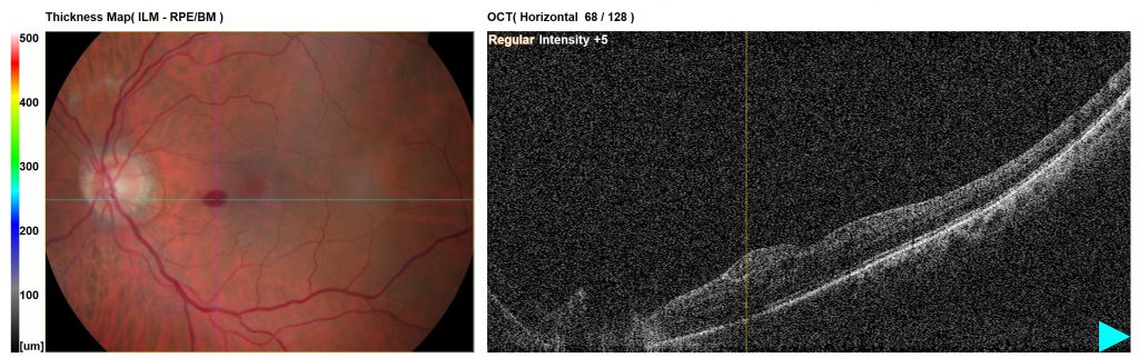
Colour photo and OCT – left macula
This is a colour photograph of the left macula with a corresponding horizontal OCT image. The colour photograph shows an oval-shaped haemorrhage nasal to the fovea, which appears to be relatively superficial as it obscures the underlying retinal vasculature and is bright red and regular in colour. The OCT scan shows some hyperreflective material underneath the inner limiting membrane (ILM), which corresponds with the location of the haemorrhage seen on the colour photograph.
Sub-inner limiting membrane haemorrhages can be due to Valsalva retinopathy (i.e. a preretinal haemorrhage caused by a sudden increase in intrathoracic or intra-abdominal pressure due to vomiting, coughing, straining or physical activity), Terson syndrome (intraocular haemorrhage associated with intracranial subarachnoid haemorrhage and increased intracranial pressure), or associated with blood dyscrasias (e.g. anaemia or haematological malignancies).
Management of sub-ILM haemorrhages depends on their location and effect on a patient’s vision. A large sub-ILM haemorrhage involving the fovea may be an indication for vitrectomy or YAG laser treatment to puncture the ILM, allowing the haemorrhage to disperse into the vitreous.
This small haemorrhage was away from the centre and was treated conservatively.