
Microcystic macular oedema secondary to ischaemic optic neuropathy
This 62-year-old man was referred to the medical retina clinic by his optician for investigation of ‘cystoid macular oedema’ following a routine sight test, which included a macular OCT scan.
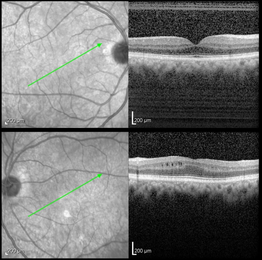
Macular OCT scan
The OCT scan of the left macula shows slit-like cystic spaces within the inner nuclear layer, inferior to the fovea.
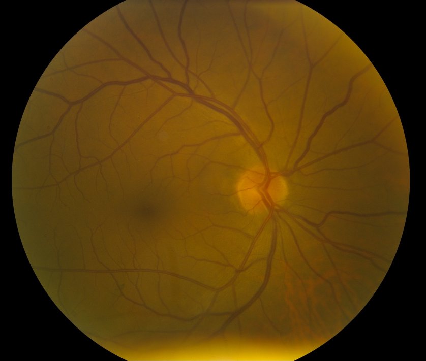
Colour fundus photograph – right eye
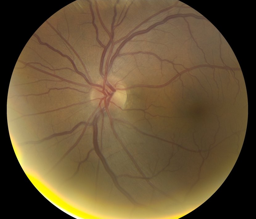
Colour fundus photograph – left eye
The left optic disc appeared slightly pale compared to the right.
This patient has a history of a left non-arteritic ischaemic optic neuropathy (NAION) with some superior visual field loss. The OCT features are typical of microcystic macular oedema , which represents retrograde retinal changes secondary to optic nerve pathology, such as demyelinating optic neuritis, glaucoma, NAION or compressive lesions, or conditions affecting the visual pathways.
When microcystic macular oedema is seen on OCT, a patient’s optic nerve function should be tested (VA, colour vision, pupil reactions, visual fields) and neuroimaging should be considered.