Central serous chorioretinopathy confirmed on FFA
This 58-year-old gentleman described a dark patch in the centre of his vision from his left eye, which had been present for approximately six months and was more noticeable under certain conditions. Visual acuities measured 6/6 right eye and 6/7.5 left.
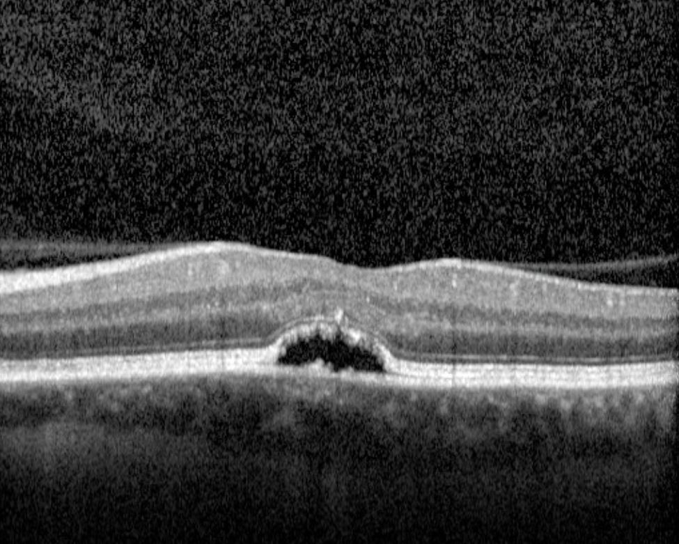
OCT scan central macula left eye
The left macular OCT scan showed some subretinal fluid at the left fovea with a slightly flattened ILM contour. The irregular ‘stalactites’ suggest chronicity. The choroidal thickness appears normal (roughly equal to the thickness of the retina) and the retinal pigment epithelium appears generally healthy in both eyes with an absence of drusen.
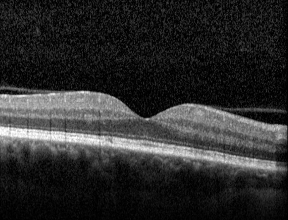
OCT scan central macula right eye
Colour fundus photograph right eye
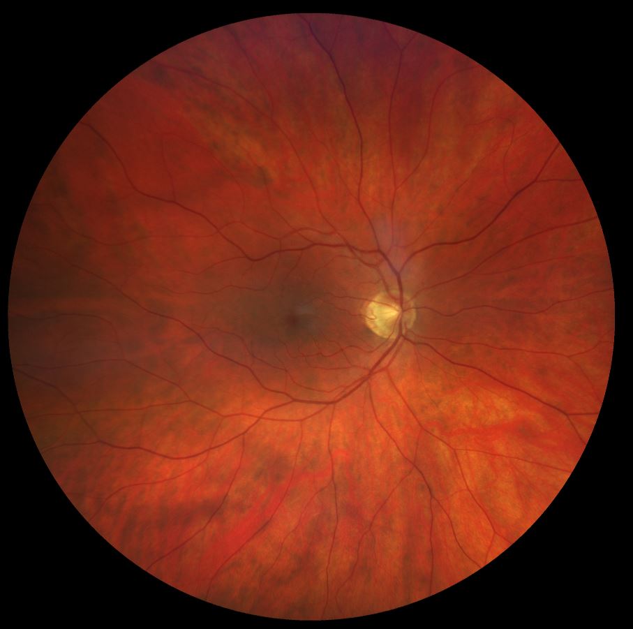
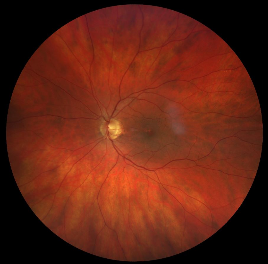
Colour fundus photograph left eye
Fundoscopy was unremarkable on the right, but there was some hypopigmentation and focal hyperpigmentation at the left fovea.
The differential diagnosis includes vitelliform maculopathy, central serous chorioretinopathy (CSCR) and age-related macular degeneration (AMD). The former two diagnoses were favoured in view of the patient’s relatively young age, the absence of drusen and mild symptoms.
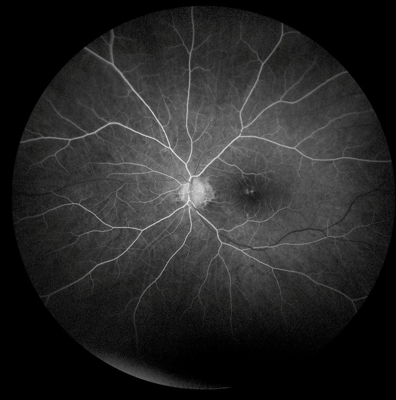
FFA left eye (early venous phase)
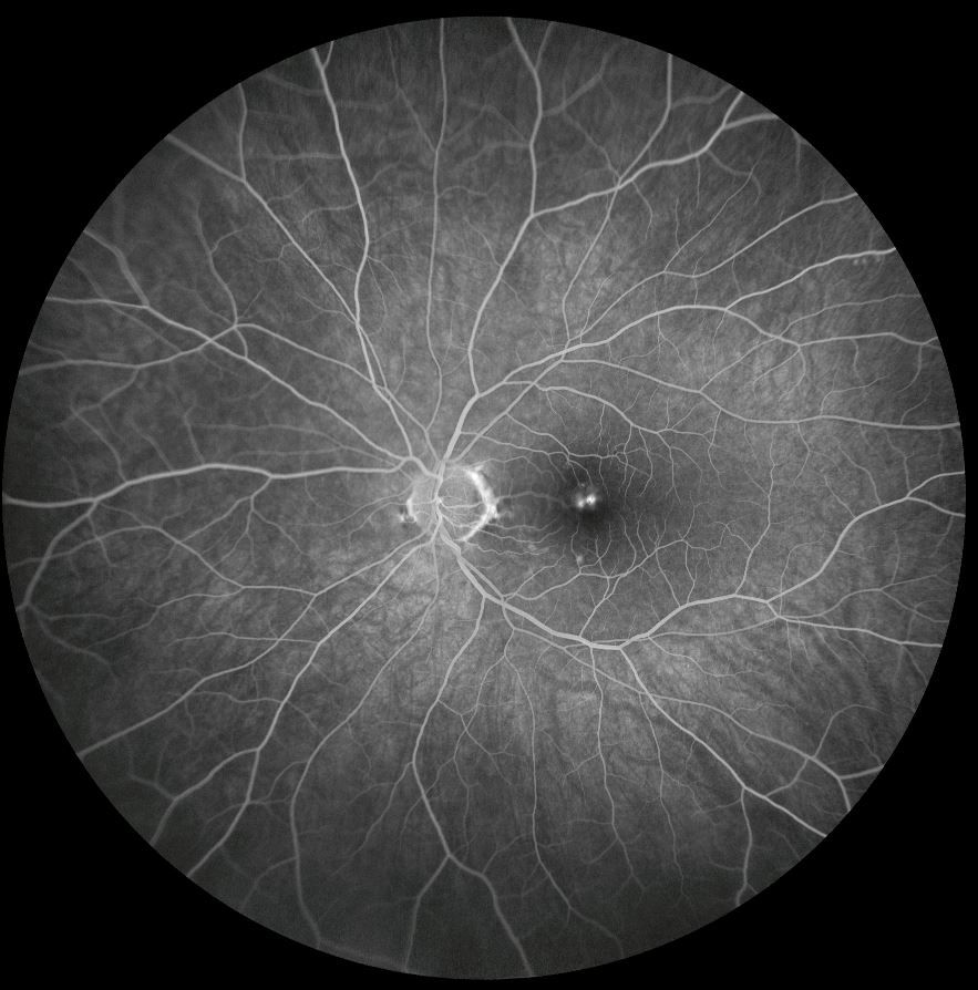
FFA left eye (arteriovenous phase)
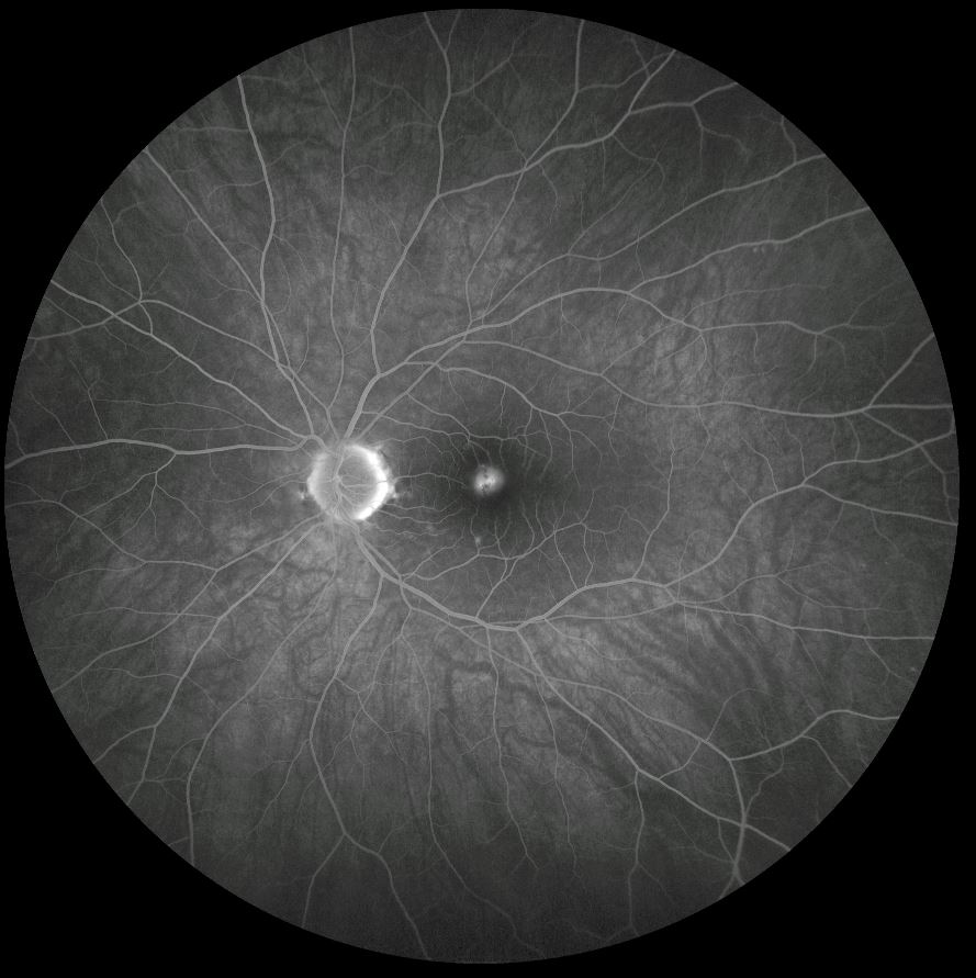
FFA left eye (late phase)
Fundus fluorescein angiography (FFA) showed two discrete areas of early hyperfluorescence at the fovea, which leaked with an ‘ink blot’ pattern later on — typical of central serous chorioretinopathy (CSCR) . Vitelliform maculopathy can show a characteristic ‘corona sign’ on FFA (hypofluorescence in the area corresponding to the vitelliform lesion, with a ring of hyperfluorescence that increases in intensity in the late phases). Fundus autofluorescence was not carried out in this case, but vitelliform lesions are usually hyperautofluorescent .
Acute CSCR is often self-limiting (usually within 3 months), but this patient was offered photodynamic therapy (PDT) in view of the duration of their symptoms.