Choroidal macrovessel presenting as an incidental OCT abnormality
A 52-year-old man was referred following his first macular OCT at a routine sight test. He was asymptomatic. The optometrist noted a raised area temporal to the fovea on OCT, with a corresponding subtle fundus change visible on colour photography that had been present on a previous photograph taken two years earlier. A choroidal osteoma was queried.
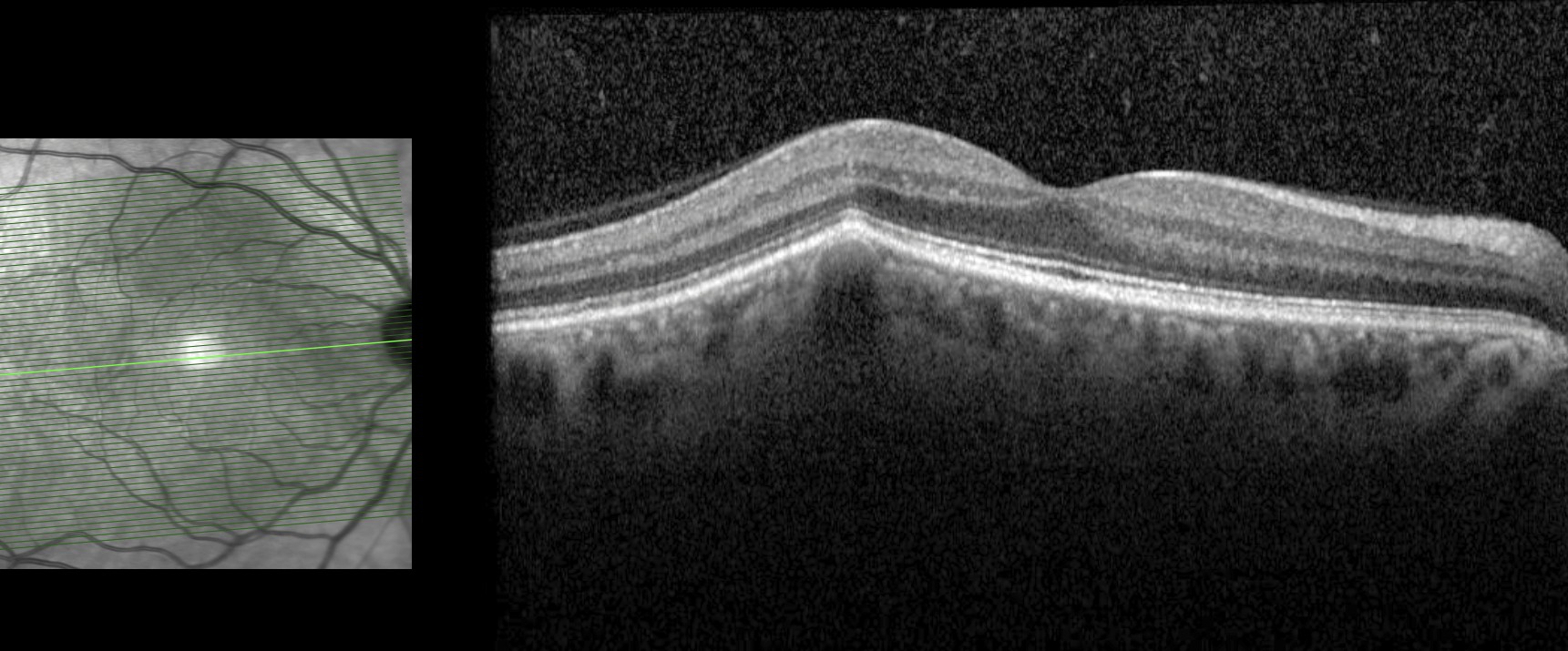
Macular OCT - right eye. Large hyporeflective space within the choroid beneath the fovea, causing elevation and abnormal contour of the overlying RPE and neurosensory retina. No subretinal or intraretinal fluid.
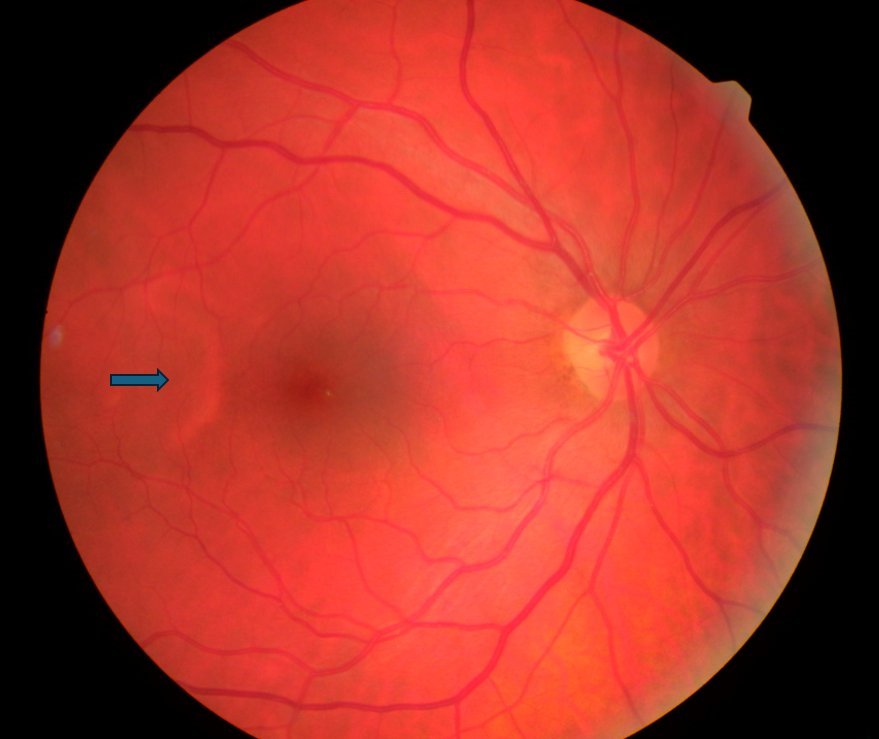
Colour fundus photograph - right eye. A dilated, tortuous choroidal vessel is visible temporal to the fovea (arrow), corresponding to the OCT abnormality.
The OCT appearances are typical of a choroidal macrovessel - a rare, anomalous dilated choroidal vessel characterised on OCT by a large hyporeflective space beneath the RPE, with posterior shadowing and elevation of the overlying retina. The corresponding tortuous vessel visible on the colour photograph temporal to the fovea confirms the diagnosis. The stable appearance over two years on fundus photography is further reassurance.
Choroidal macrovessels are benign incidental findings that do not require treatment in the absence of complications such as subretinal fluid or neovascularisation. They can mimic more sinister pathology - including choroidal tumours and choroidal granulomas - making multimodal imaging important in establishing the diagnosis. The condition is well described in the literature, including a multimodal imaging series by Gallo, de Silva et al. (BJO 2022) which characterised the range of fundus photography, OCT, and angiographic appearances across 16 eyes.
This case illustrates the value of optometrists submitting good quality imaging with their referrals - it allowed confident remote diagnosis, facilitated co-management with the patient remaining under routine optometric care without a hospital visit, and provided an opportunity for discussion and education that both parties found rewarding.