A growing choroidal lesion: the importance of serial imaging
A 57-year-old woman was referred urgently with a two-month history of reduced left vision and a hypopigmented raised lesion within the left macula that had increased in size compared to fundus photographs and OCT from July 2024. Visual acuity measured 6/6 right and 6/18 left. Intraocular pressures were normal bilaterally. The left acuity corrected to 6/6 with a plus lens - consistent with a hypermetropic shift induced by the growing choroidal lesion effectively shortening the axial length. Colour photographs were not submitted with the referral.
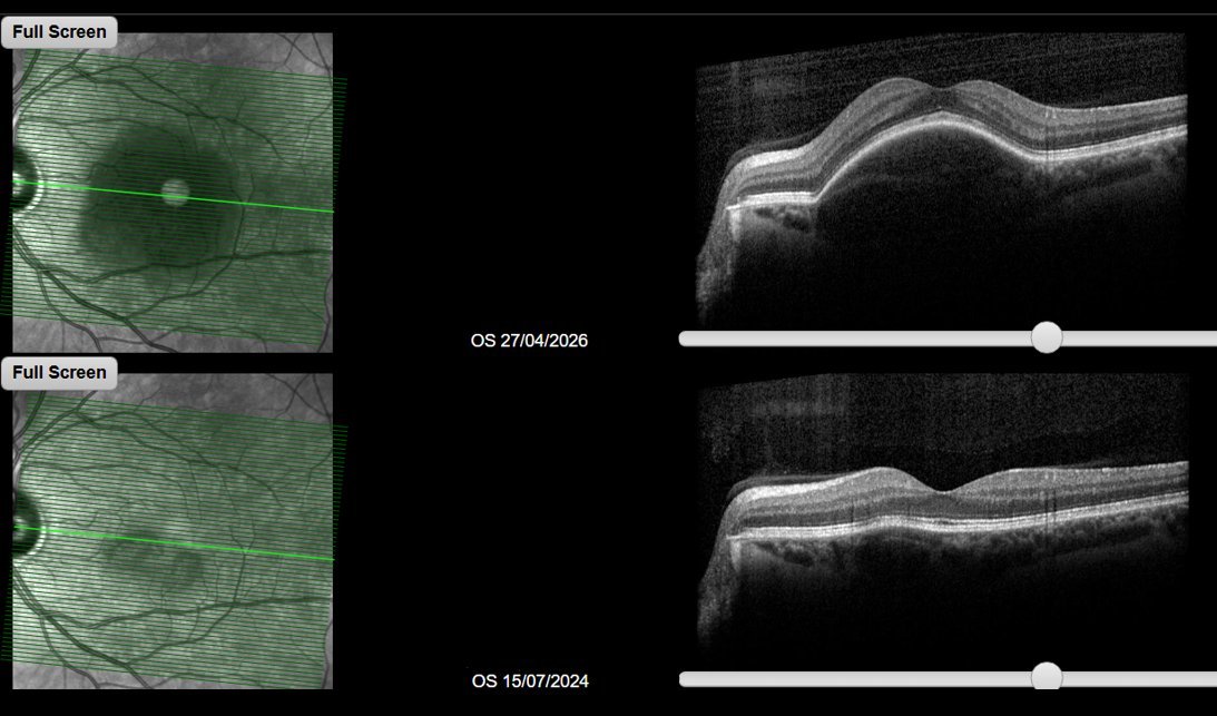
Comparative macular OCT - left eye. Upper scan (April 2026) and lower scan (July 2024) showing interval growth of a dome-shaped RPE elevation with an underlying hyporeflective choroidal thickening, consistent with a serous pigment epithelial detachment overlying an expanding choroidal lesion.
The OCT demonstrates a dome-shaped elevation of the RPE, associated with a serous pigment epithelial detachment (PED) and hyporeflective thickening of the underlying choroid. The lesion has grown appreciably between the two timepoints.
This appearance is consistent with a choroidal mass causing secondary elevation of the overlying RPE, and circumscribed choroidal haemangioma is on the differential - a benign vascular hamartoma that typically presents in the fourth to sixth decade with progressive visual symptoms from associated exudation. A pachychoroid-related serous PED in the context of CSCR spectrum disease is also possible, though the documented growth over two years and the patient’s age and sex make this less typical. However, the differential diagnosis must include choroidal melanoma and choroidal metastasis, both of which can produce a similar OCT appearance. Colour fundus photography was not available with this referral; the characteristic orange-red colour of a haemangioma, if present, would be a useful clinical pointer, and its absence from the submitted images is a limitation.
Formal assessment including colour fundus photography, B-scan ultrasonography, and fluorescein and indocyanine green angiography will be needed to characterise the lesion. This patient will be seen in a medical retina clinic within six weeks. This case illustrates the value of serial imaging in optometric practice - the ability to demonstrate interval growth transformed an incidental finding into an urgent and clearly justified referral.