Two incidental findings, one routine sight test
A 64-year-old woman was referred following an abnormal OCT appearance in the right eye at a routine sight test. She was entirely asymptomatic with good visual acuity bilaterally.
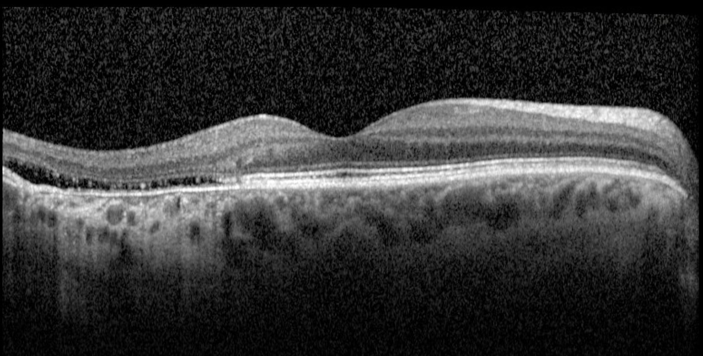
Macular OCT - right eye. A focal area of outer retinal loss temporal to the fovea with cavitation of the outer retinal layers producing characteristic hanging projections (“stalactites”) from the ellipsoid zone into the subretinal space. The RPE and photoreceptor layer are absent in the affected area, with Bruch’s membrane intact and a corresponding hypertransmission defect into the choroid. Foveal architecture is preserved.
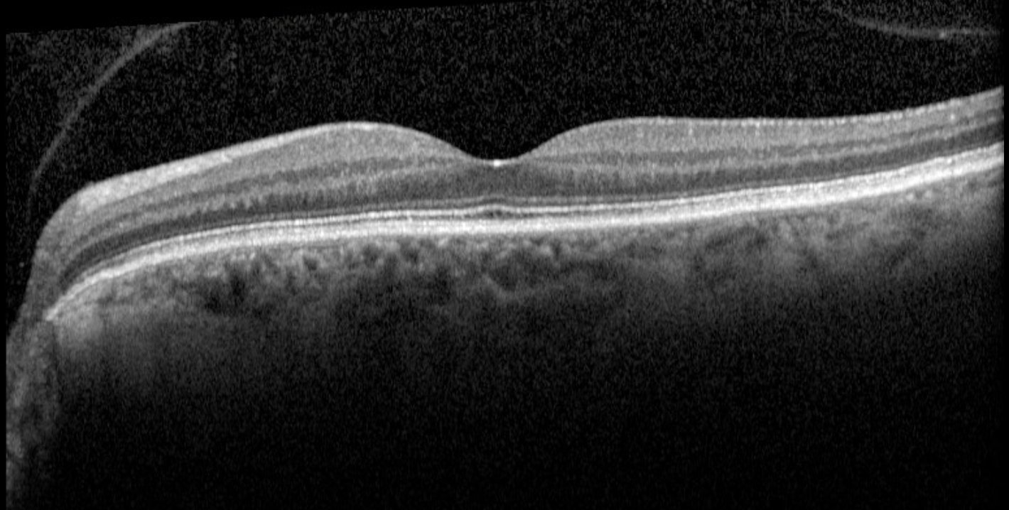
Macular OCT - left eye. Normal foveal contour. Posterior vitreous detachment noted. No macular pathology.
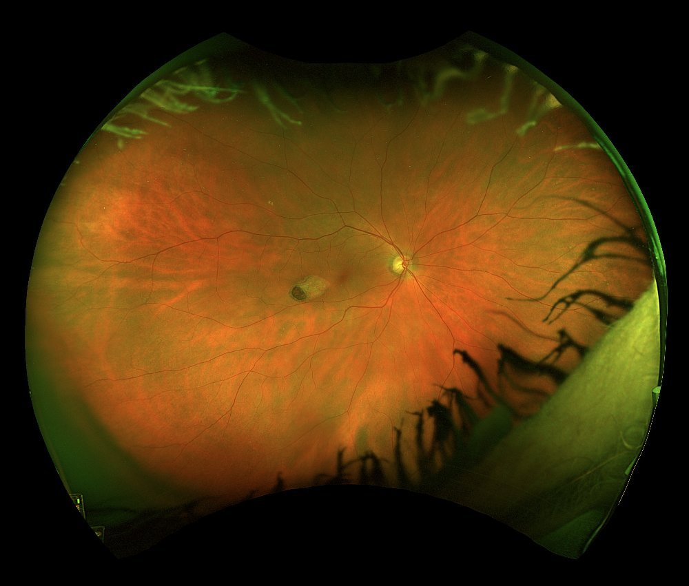
Widefield colour fundus photograph - right eye. A characteristic torpedo-shaped hypopigmented lesion is visible in the temporal macula along the horizontal raphe, with its pointed tip directed towards the fovea.
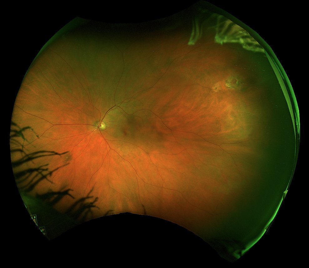
Widefield colour fundus photograph - left eye. Normal macula. Incidental longstanding retinal tear in the superotemporal periphery with surrounding reactive pigmentation.
The right OCT and fundus appearances are diagnostic of torpedo maculopathy - a rare, benign congenital anomaly of the RPE first described by Roseman and Gass in 1992. The lesion arises along the horizontal raphe temporal to the fovea, with its characteristic torpedo shape and tip pointing towards the central macula. On OCT, the outer retinal cavitation with hanging projections from the ellipsoid zone represents a Type 2 lesion in the OCT subtype classification proposed by Wong et al. (Clin Experiment Ophthalmol 2015) , which divided torpedo maculopathy into Type 1 (outer retinal attenuation without cavitation) and Type 2 (outer retinal attenuation with cavitation). The intact Bruch’s membrane and preserved foveal architecture are reassuring features. The condition is typically stationary and, as here, discovered incidentally in adulthood despite being congenital. Rare complications include choroidal neovascularisation, which should prompt referral if new symptoms develop.
The left widefield photograph revealed an incidental retinal tear in the superotemporal periphery, surrounded by reactive pigmentation consistent with a stable, chronic lesion. In an asymptomatic patient with no history of recent floaters or flashes, no treatment is required.
Both findings were benign and required no active intervention. This patient will be monitored with periodic optometric review and has been advised to report promptly if any new visual symptoms develop.