Blood pressure 260/150: what the fundus revealed
A 55-year-old man was referred for ophthalmology review by the medical team during an inpatient admission. He had no previous ocular or medical history and was visually asymptomatic. Visual acuity was 6/6 bilaterally and intraocular pressures were normal. He had been admitted to the intensive care unit three days earlier with shortness of breath and fatigue following a recent respiratory infection, and was found to have a blood pressure of 260/150 mmHg. Investigations revealed acute kidney injury with a creatinine of 500, echocardiography showing severely impaired left ventricular function with an ejection fraction of 15-20%, and subsequent renal biopsy confirming hypertensive changes with chronic damage. He was commenced on multiple antihypertensive agents and transferred to the renal ward.
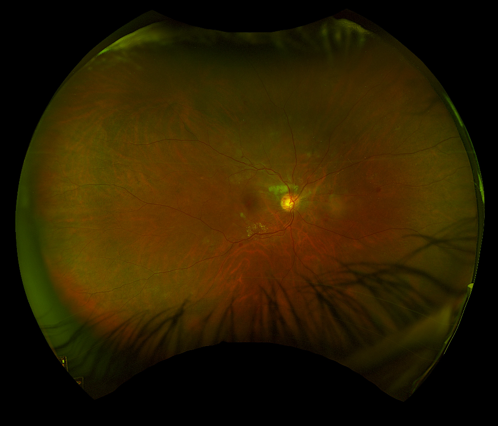
Widefield colour fundus photograph - right eye. Peripapillary cotton wool spots, flame-shaped haemorrhages, disc collaterals, circinate hard exudates along the inferior arcade, and arteriolar attenuation.
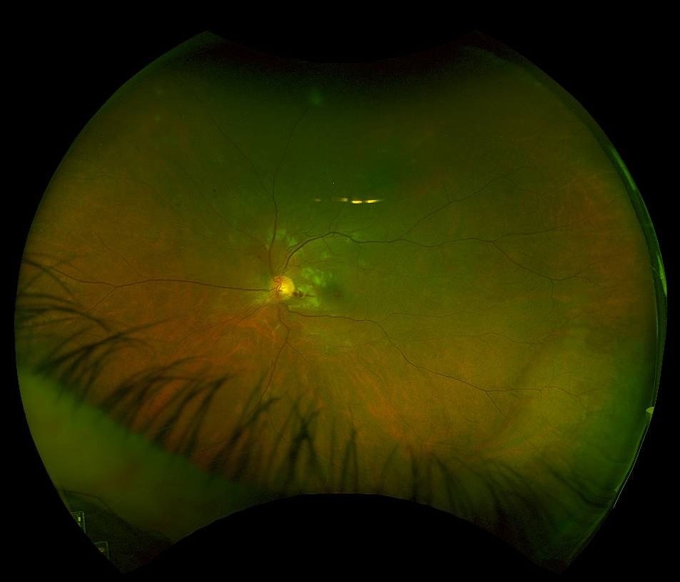
Widefield colour fundus photograph - left eye. Peripapillary cotton wool spots, flame-shaped haemorrhages, disc collaterals, and arteriolar attenuation. The striking peripapillary distribution of cotton wool spots and haemorrhages reflects acute nerve fibre layer ischaemia from the hypertensive insult.
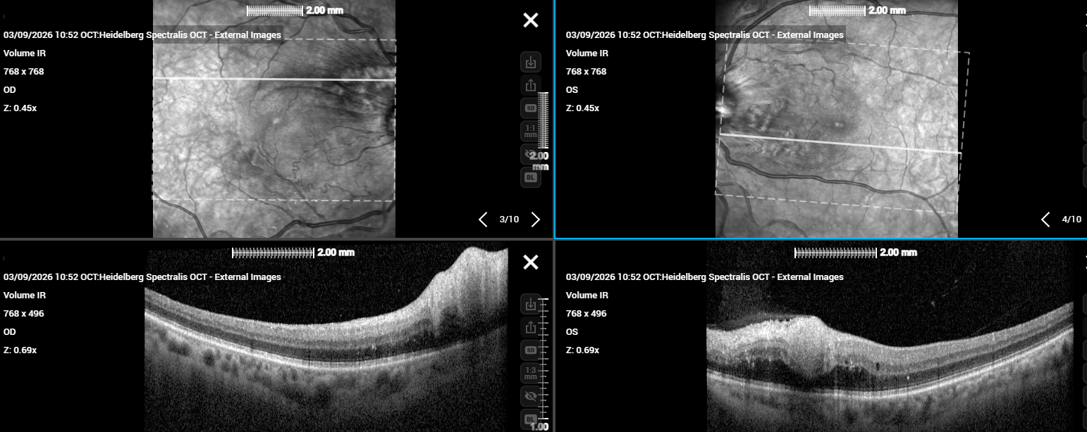
Bilateral macular OCT. Right eye: hyperreflective thickening spreading through multiple inner retinal layers at several locations, consistent with nerve fibre layer infarction - the OCT correlate of cotton wool spots. Left eye: similar inner retinal hyperreflectivity with associated intraretinal fluid.
Bilateral fundoscopy revealed peripapillary cotton wool spots , flame-shaped haemorrhages, disc collaterals, arteriolar attenuation, and circinate hard exudates along the inferior arcade of the right eye - the full picture of severe hypertensive retinopathy . Cotton wool spots represent focal nerve fibre layer infarcts caused by pre-capillary arteriolar occlusion in the context of acute, severe elevation in blood pressure. On OCT they appear as hyperreflective thickening spreading through multiple inner retinal layers - superficially similar to other inner retinal lesions such as retinal astrocytic hamartomas, which project from the nerve fibre layer surface (as illustrated in the tuberous sclerosis case on this site) - but the clinical context, acute onset, and fundoscopic appearance of fluffy white peripapillary patches distinguish them immediately. Unlike hamartomas, which are stable congenital lesions, cotton wool spots resolve spontaneously within six to twelve weeks as the ischaemic insult declares itself and the axoplasmic debris is cleared.
Using the Wong and Mitchell classification - still the most widely used system, originally proposed in 2004 - this patient has severe hypertensive retinopathy, defined as moderate retinopathy (haemorrhages, cotton wool spots, hard exudates) with optic disc involvement. No papilloedema was present, which places this at the upper end of the moderate category rather than malignant retinopathy, though the degree of systemic end-organ damage - cardiac, renal, and retinal - was profound.
He was reassured that he was visually asymptomatic and that his blood pressure was now under control, and has been booked for medical retina clinic review at two months. This case is a reminder that retinal findings detected incidentally in otherwise asymptomatic patients - most often by their optometrist - can represent the first presentation of serious systemic disease. Hypertensive retinopathy, haematological malignancies, and other systemic conditions may all declare themselves at the slit lamp or on fundoscopy before any other symptoms have emerged. The retina is a window to the circulation, and the optometrist who identifies these changes may be the first clinician to avert a life-threatening emergency.